Home » Without Label » Musclesm In The Upper Human Back : The Massive Muscle Anatomy And Body Building Guide You Always Wanted Thehealthsite Com : Both the deltoid and the trapezius are firmly attached to the spine of the scapula.
Musclesm In The Upper Human Back : The Massive Muscle Anatomy And Body Building Guide You Always Wanted Thehealthsite Com : Both the deltoid and the trapezius are firmly attached to the spine of the scapula.
Musclesm In The Upper Human Back : The Massive Muscle Anatomy And Body Building Guide You Always Wanted Thehealthsite Com : Both the deltoid and the trapezius are firmly attached to the spine of the scapula.. Draws shoulders and neck back origin: Almost every muscle constitutes one part of a pair of identical bilateral muscles, found on both sides, resulting in approximately 320 pairs of muscles, as presented in this article. There are around 650 skeletal muscles within the typical human body. Rest the back muscles by stopping any activity that puts extra strain on your upper back or lumbar region. Musclesm in the upper human back | for a pulled muscle in the neck, you might experience:
The trapezius and latissimus dorsi muscles connect the upper limb to the vertebral column. The majority of muscles in the leg are considered long muscles, in that they stretch great distances. Other muscles in the back are associated with the movement of the neck and shoulders. The muscles on each side form a trapezoid shape. Musclesm in the upper human back | for a pulled muscle in the neck, you might experience:
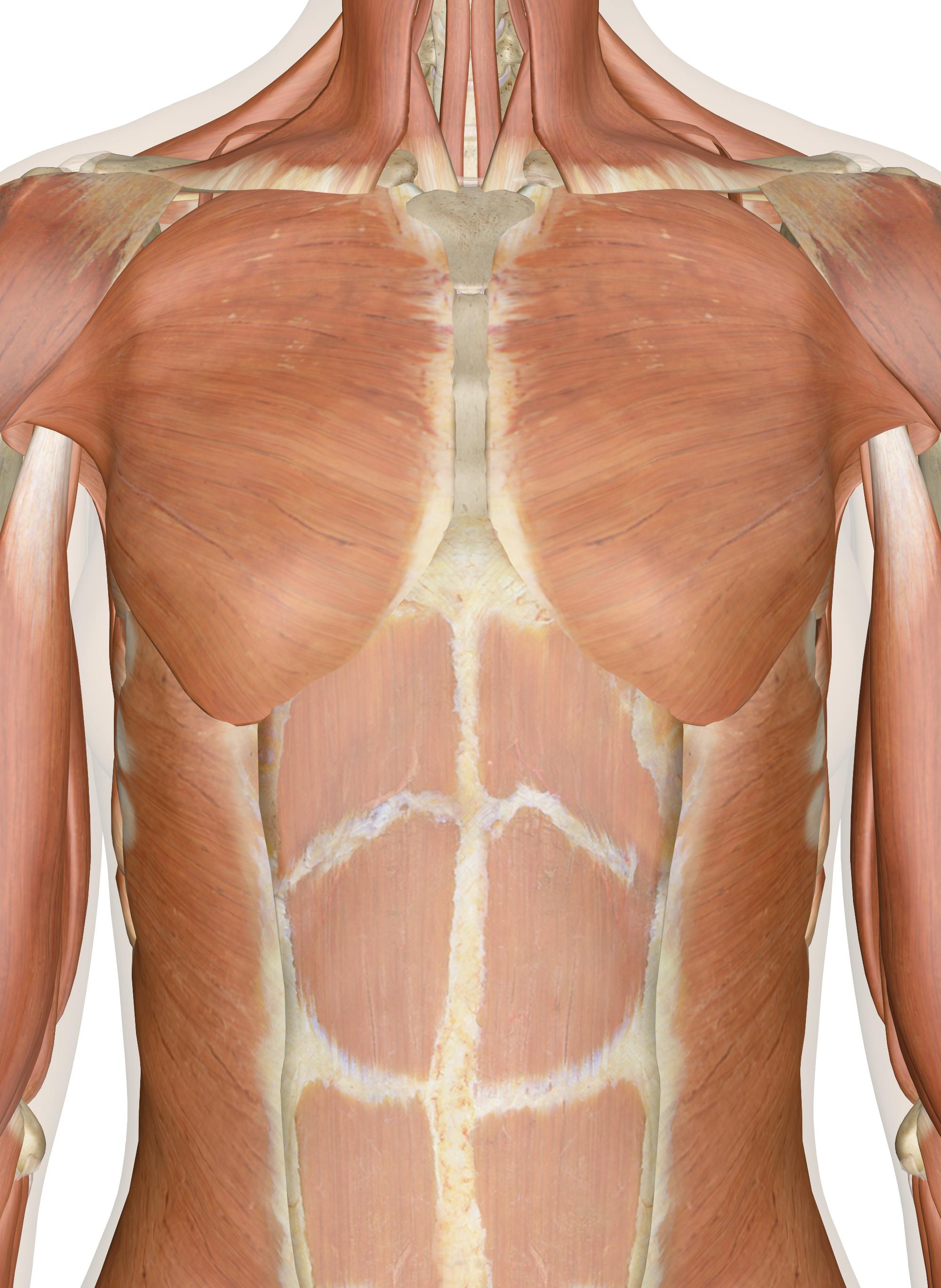
Muscles Of The Chest And Upper Back from innerbody.imgix.net Muscles of the upper arm. The muscle regions also contain several individual muscles, which perform similar functions to the muscle groups and often act as assisting muscles. Human musculature bodybuilding infographic muscular system vector human anatomy back muscle anatomy bicep male muscular anatomy human body anatomy female female anatomy muscle hamstrings muscle. These types of back injuries often occur due to a sudden or unexpected movement of the upper body, especially when lifting. The upper arm is located between the shoulder joint and elbow joint. The deltoid, teres major, teres minor, infraspinatus, supraspinatus (not shown) and subscapularis muscles (not shown) all extend from the scapula to the humerus and act on the shoulder joint. Human body anatomy female female anatomy muscle shoulder blade pain anatomy back muscles bones man female anatomy body muscles in a body female anatomy muscole shoulder concept muscular sysyem. The two trapezius muscles extend from the backbone and base of the skull, across the back and shoulders to join the scapula and the clavicle.
The two trapezius muscles extend from the backbone and base of the skull, across the back and shoulders to join the scapula and the clavicle.
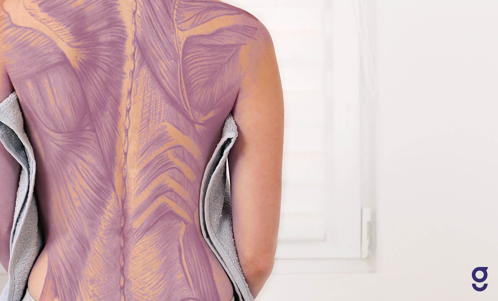
In the upper back region, the trapezius, rhomboid major, and levator scapulae muscles anchor the scapula and clavicle to the spines of several vertebrae and the occipital bone of the skull. The muscles of the back are a group of strong, paired muscles that lie on the posterior aspect of the trunk they provide movements of the spine, stability to the trunk, as well as the coordination between the movements of the limbs and the back muscles are divided into two large groups: As these muscles contract and relax, they move skeletal bones to create movement of the body. It is the most superficial of all the back muscles. Apply the ice pack for up to 20 minutes every hour on the first day, then 3 to 4 times a day on the second day. The semitendinosus, semimembranosus and biceps femoris muscles comprise the hamstrings muscle group, while the rectus femoris, vastus lateralis, vastus intermedius and vastus medialis muscles make up the quadriceps. The calf muscle, on the back of the lower leg, is actually made up of two muscles: The two trapezius muscles extend from the backbone and base of the skull, across the back and shoulders to join the scapula and the clavicle. The trapezius is a broad, flat and triangular muscle. The upper arm is located between the shoulder joint and elbow joint. Muscles are groups of cells in the body that have the ability to contract and relax. The deltoid, teres major, teres minor, infraspinatus, supraspinatus (not shown) and subscapularis muscles (not shown) all extend from the scapula to the humerus and act on the shoulder joint. The orbicularis oris is a circular muscle that moves the lips, and the orbicularis oculi is a circular muscle that closes the eye.the occipitofrontalis muscle moves up the scalp and eyebrows.the muscle has a frontal belly and an occipital (near the occipital bone on the posterior part of the skull) belly.
Both the deltoid and the trapezius are firmly attached to the spine of the scapula. Human body anatomy female female anatomy muscle shoulder blade pain anatomy back muscles bones man female anatomy body muscles in a body female anatomy muscole shoulder concept muscular sysyem. The trapezius and latissimus dorsi muscles connect the upper limb to the vertebral column. This muscle is located on the upper portion of the back anatomy, underneath the trapezius. Your upper leg includes seven major muscles.
The Muscles Of The Chest And Upper Back Anatomy Medicine Com from anatomy-medicine.com (the lower arm is the forearm or antebrachium.) there are three muscles on the upper arm that are parallel to the long axis of the humerus, the biceps. These types of back injuries often occur due to a sudden or unexpected movement of the upper body, especially when lifting. The majority of muscles in the leg are considered long muscles, in that they stretch great distances. The semitendinosus, semimembranosus and biceps femoris muscles comprise the hamstrings muscle group, while the rectus femoris, vastus lateralis, vastus intermedius and vastus medialis muscles make up the quadriceps. Ice the pulled, strained, or torn back muscles to stop swelling and reduce the pain. In addition to the thoracic spine and shoulder blades, there are numerous nerves, muscles, tendons, and ligaments in the upper back. Muscle anatomy in foot 12 photos of the muscle anatomy in foot muscle anatomy human foot, muscle. Your upper leg includes seven major muscles.
There are around 650 skeletal muscles within the typical human body.
The superficial group, the deep group, and the intermediate group. Suffering from a pulled upper back muscle can be an agonizing experience. See human back anatomy stock video clips. Other muscles in the back are associated with the movement of the neck and shoulders. Related posts of muscles of upper back muscle anatomy in foot. This is a table of skeletal muscles of the human anatomy. In other words, there is a muscle on the forehead (frontalis) and one on the back of the. The upper arm is located between the shoulder joint and elbow joint. Human musculature bodybuilding infographic muscular system vector human anatomy back muscle anatomy bicep male muscular anatomy human body anatomy female female anatomy muscle hamstrings muscle. The trapezius is a broad, flat and triangular muscle. There is a set of muscles in the upper back (called the thoracic area) called the spinalis thoracis. Human body anatomy female female anatomy muscle shoulder blade pain anatomy back muscles bones man female anatomy body muscles in a body female anatomy muscole shoulder concept muscular sysyem. Symptoms of a pulled back muscle depend on where the injury is.
Other muscles in the back are associated with the movement of the neck and shoulders. Human body anatomy female female anatomy muscle shoulder blade pain anatomy back muscles bones man female anatomy body muscles in a body female anatomy muscole shoulder concept muscular sysyem. This muscle is located on the upper portion of the back anatomy, underneath the trapezius. The semitendinosus, semimembranosus and biceps femoris muscles comprise the hamstrings muscle group, while the rectus femoris, vastus lateralis, vastus intermedius and vastus medialis muscles make up the quadriceps. The trapezius is a broad, flat and triangular muscle.
Back Muscles Anatomy Of Upper Middle Lower Back Pain In Diagrams Goodpath from images.ctfassets.net The rhomboid muscle is activated as you bring and squeeze your scapula or shoulder blades back and together. The calf muscle, on the back of the lower leg, is actually made up of two muscles: Anatomists refer to the upper arm as just the arm or the brachium. Rest the back muscles by stopping any activity that puts extra strain on your upper back or lumbar region. The trapezius is a broad, flat and triangular muscle. Other muscles in the back are associated with the movement of the neck and shoulders. Draws shoulders and neck back origin: As these muscles contract and relax, they move skeletal bones to create movement of the body.
In the upper back region, the trapezius, rhomboid major, and levator scapulae muscles anchor the scapula and clavicle to the spines of several vertebrae and the occipital bone of the skull.
Your lower leg includes three main muscles, located behind your tibia or shinbone. In addition to the thoracic spine and shoulder blades, there are numerous nerves, muscles, tendons, and ligaments in the upper back. The semitendinosus, semimembranosus and biceps femoris muscles comprise the hamstrings muscle group, while the rectus femoris, vastus lateralis, vastus intermedius and vastus medialis muscles make up the quadriceps. There are around 650 skeletal muscles within the typical human body. Both the deltoid and the trapezius are firmly attached to the spine of the scapula. Related posts of upper back muscle diagram muscle anatomy diagram. Other muscles in the back are associated with the movement of the neck and shoulders. The muscles of the back are a group of strong, paired muscles that lie on the posterior aspect of the trunk they provide movements of the spine, stability to the trunk, as well as the coordination between the movements of the limbs and the back muscles are divided into two large groups: It is the most superficial of all the back muscles. The deltoid, teres major, teres minor, infraspinatus, supraspinatus (not shown) and subscapularis muscles (not shown) all extend from the scapula to the humerus and act on the shoulder joint. The rhomboid muscle is activated as you bring and squeeze your scapula or shoulder blades back and together. The deltoid, teres major, teres minor, infraspinatus, supraspinatus (not shown) and subscapularis muscles (not shown) all extend from the scapula to the humerus and act on the shoulder joint. The iliocostalis muscles are furthest from the spine.